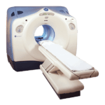
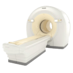
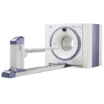
It is now possible to use PET/CT scans to determine the presence or absence of coronary artery calcium in patients with chest pain
According to a study presented last month at the American College of Cardiology (ACC) 2019 meeting in New Orleans, it is now possible to use PET/CT scans to determine the presence or absence of coronary artery calcium in patients with chest pain. This can help determine which individuals have an increased risk of a future major adverse cardiovascular event, and have the greatest need for quick remedial action.
The researchers, who were from the Intermountain Medical Center Heart Institute in Salt Lake City, discovered that when coronary artery calcium was present in patients, they were more likely to undergo revascularization or coronary angiography with 90 days of scanning and have high-grade obstructive coronary artery disease. Patients who had no evidence of calcification could also be better directed to a more appropriate noncardiac follow-up, as suggested by these findings.
As stated by principal investigator Viet Le, a physician’s assistant and researcher at Intermountain: “We are interested in coronary artery calcium from a primary prevention and risk reduction standpoint. It followed that with the imaging that we do at Intermountain, the question arose: Is there a better way in those folks who have not had coronary artery disease, but show with symptoms, to better stratify who should have a stress test and who should undergo a workup for something else?”
Going to the E.R. for chest pain is common amongst Americans, with 6 million people going annually, while 3.8 million of them (68%) go on to have stress tests or have imaging done. As to be expected, there is a great number of these patients who do not have a positive test result for myocardial infarction or other coronary event at all, so hospital resources are not being enacted to the best of their ability. Dr. Le continued: “That is a lot of money for the healthcare system and for the patient when perhaps there is a better way to improve which population we stress test.”
Because a lack of positive test result is often the case, PET/CT could offer a noninvasive option to determine the degree of coronary artery calcium, the deposits of which suggest coronary artery disease and/or a future major adverse cardiovascular event. It should be noted, however, that it is unclear how well coronary artery calcium predicts risk for major cardiovascular events.
In order to remedy the uncertainty somewhat, Dr. Le and his colleagues identified 5,547 patients with no history of coronary artery disease who came to Intermountain Heart Institute from April 2013 to June 2016 with complaints of chest pain. The patients were given rubidium-82 PET/CT scans, to test for ischemia, which disrupts normal blood flow through the heart’s arteries to its muscle tissue; as well as to determine the presence of coronary artery calcium deposits on the walls of the arteries, which would indicate atherosclerosis or plaque.
Get Started
Request Pricing Today!
We’re here to help! Simply fill out the form to tell us a bit about your project. We’ll contact you to set up a conversation so we can discuss how we can best meet your needs. Thank you for considering us!
Great support & services
Save time and energy
Peace of mind
Risk reduction
Within the first 90 days after the scans, researchers visually took note of the absence and presence of coronary artery calcium and correlated those findings with patient outcomes relative to coronary angiography, high-grade coronary artery disease, and revascularization. After 90 days but less than 4 years later, the researchers tracked major adverse cardiovascular events (such as death, hospitalization, or myocardial infarction), as well as revascularization.
The end results of the study showed that researchers found that 45% of patients (2,515) had coronary artery calcium, while 55% showed no signs at all (3,302 individuals). Once they looked at the results of the two groups, they discovered that the patients with the coronary artery calcium fared poorly in comparison to the patients with no calcium present.
The results from the study suggested to researchers that a “coronary artery calcium first” approach would be most beneficial to separate symptomatic patients who are prime candidates for additional evaluation (including further functional stress tests) from patients who only have a small risk of coronary artery disease or future major adverse event. These patients should be referred to a noncardiac follow-up to diagnose the cause of their chest pain.
According to Dr. Le, “When not stuck on coronary artery calcium, we could assess for maybe pulmonary, musculoskeletal, or gastrointestinal [causes], but we would be pretty darn sure it is not coronary-related. On the other hand, if the patient has a calcium score greater than 0, these are folks that you can immediately send through to have a PET scan.”
Once hospitals are better able to direct their patients to a more suitable test or scan, patient throughput would be greatly enhanced because emergency departments “would have eliminated, say, 50% of those folks who would’ve sat on your schedule. It would improve those emergency department times in terms of patient turnaround and improve the efficiency of the system.”
Le continued that Intermountain clinicians have begun building a research protocol to “put our money where our mouth is, so to speak. We are prospectively checking to see if a coronary artery calcification as a first strategy will indeed identify folks who do not need to have a PET/CT but are better served by being met earlier by a noncardiac physician. It is just a hypothesis at this point, but it is an observation with a real-world finding. Now we have to test it going forward, and we will.”


