Learn about C-Arm image acquisition and how it works
Image acquisition on a mobile C-arm for fluoroscopy procedures is either done via an image intensifier (ii) or a flat plate detector.
Early models of stationary flouro units were either mounted to the floor or the ceiling and couldn’t achieve extreme angles. They were used in cardiac catheterization labs for angiograms or interventional radiology suites for vascular peripheral studies. But stationary units lacked the maneuverability to get extreme angles of bony and vascular anatomy. Therefore, a more mobile x-ray unit was developed that could be moved from room to room and angled around the patient without obstructing the surgical site.
A mobile C-arm unit is developed for Surgery
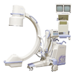
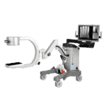
Philips came out with the first mobile C-arm that used fluoroscopy for surgical ortho work in 1955. The C-arm housed an x-ray generator/tube with an image intensifier mounted on the opposite end of a half-moon arm. The fluoroscopic mobile C-arm was attached to a carriage which allowed easy transport from room to room in the OR suite. The c-arm could move around and under the patient and achieve extreme angles for various surgical procedures. The image intensifier attached to the other end of the C-arm brightened the image without the need for increased radiation.
Fluoroscopy: The imaging of live x-ray images
Fluoroscopic C -arms come with two options for image reconstruction: The image intensifier and the flat plate detector.
Before the Image Intensifier, Thomas Edison, in the 1890’s developed a phosphorescent screen to view live x-ray images of patients swallowing barium and other contrast medium. During an upper gastrointestinal (Ugi) study, the contrast was seen flowing through the esophagus into the stomach on a phosphorescent screen.
This phosphorescent or “flouro” screen was coated with calcium tungstate to produce a brighter x-ray image that showed the live images of the internal anatomy. The images were very dim and unclear. Radiologists had to sit in darkened rooms and look directly into the x-ray beam to visualize the obscure images of internal organs.
These studies were done without lead shielding and required heavy exposure to radiation. Many Radiologists and other early 19th century practitioners suffered from cataracts and other forms of radiation induced cancers.
Get Started
Request Pricing Today!
We’re here to help! Simply fill out the form to tell us a bit about your project. We’ll contact you to set up a conversation so we can discuss how we can best meet your needs. Thank you for considering us!
Great support & services
Save time and energy
Peace of mind
Risk reduction
The Image Intensifier improves image quality
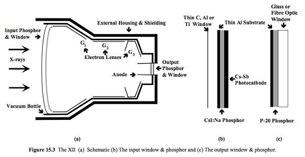
The image intensifier was developed to reduce radiation exposure and to increase the brightness of the fluoroscopic image. The x-ray photons pass through the patient into the input phosphor layer of the II and converts them to light photons. These light photons pass through a series of electron lenses that amplify the light signal or “intensifies” the light, before passing through the output phosphor. The output phosphor is coupled to a camera where the image is projected on a monitor in the room.
Essentially, the II takes “low light” x-ray photons (input phosphor) and converts them to high light photons (output phosphor). These light photons are processed by computer algorithms to produce an image projected on a monitor.
Digital Fluoroscopic C-arms use flat plate detectors to produce digital images
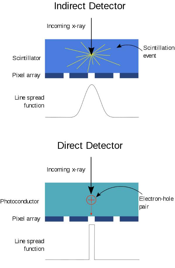
The flat plate detector uses a cesium iodide (CsI) scintillation layer coated onto the amorphous silicon photodiodes. The photodiodes capture the visible light signal to form the digital image.
The x-rays pass through the CsI layer converting the x-rays to light quanta. The quanta pass through a “film-thin” layer of photodiodes and transistors to produce a digital image. Much like cameras on smart phones, digital images can be stored, printed, or wirelessly transferred to picture archival systems.
Flat plate detectors are thin and can easily fit into tight spaces in the OR during hip pinnings and femur roddings. Digital Flat Detectors use less radiation to produce a diagnosable image and eliminate pincushion and vignetting distortion.
Image intensifiers and flat plate detectors on C-arms convert x-ray photons to light which are then converted to a video signal. The image is viewed on a TV monitor or wirelessly transferred to an image archival system.
The differences between the II and FPD is the design, and the way X-ray images are reconstructed. Image Intensifiers use a large vacuum sealed tube to produce live images. Whereas FPD uses a layer of CsI phosphor over an array of photodiodes to capture a live digital flouro image. On both models, the images are viewed on a TV monitor and stored in a picture archival system.
Sources:
https://radiologykey.com/image-intensified-fluoroscopy/
https://www.imagewisely.org/Imaging-Modalities/Fluoroscopy/Modern-Imaging-Systems
https://en.wikipedia.org/wiki/Fluoroscopy
Images from wikipedia.org

