PTSD is defined as an anxiety disorder that is caused when a person experiences a traumatic event
PTSD is defined as an anxiety disorder that is caused when a person experiences a traumatic event resulting in serious physical injury or with the potential to cause serious physical injury. Up to 6.8 percent of U.S. adults will experience the disorder in their lifetime.
Living with PTSD can be incredibly difficult. Sufferers can have strong memories of the event creep up on them, horrific nightmares, emotional numbness, intense guilt or worry, sudden anger outburst, feeling “on edge,” and avoiding thought and situations that remind them of the event.
Recent MRI imaging studies have shown that there is a surprising difference in brain structure among adults with PTSD that survived an earthquake and adult survivors without PTSD. In the study, the researchers wanted to explore the relationships of abnormalities and the clinical severity and duration of time since the trauma.
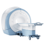
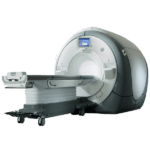
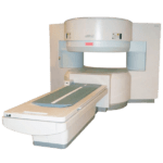
The study’s senior author is Qiyong Gong M.D., Ph.D., from Huaxi MR Research Center at West China Hospital of Sichuan University in Chengdu, China. He says that it was important for the study to compare the PTSD patients with individuals who had experienced the same trauma in order to gain information about the brain altercations that took place with the PTSD patients that are different and occur above and beyond general stress responses. Ultimately, there were 67 PTSD patients and 78 non PTSD patients, and they were all scanned with a 3.0T MRI.
Get Started
Request Pricing Today!
We’re here to help! Simply fill out the form to tell us a bit about your project. We’ll contact you to set up a conversation so we can discuss how we can best meet your needs. Thank you for considering us!
Great support & services
Save time and energy
Peace of mind
Risk reduction
Using the Clinician Administered PTSD Scale (CAPS), the patients were evaluated by trained earthquake support psychologists, and those with a CAP score of >50 were further evaluated to determine if the patient had PTSD.
At the end of the study, it was revealed that the PTSD patients had greater cortical thickness in certain brain regions, as well as reduced volume on other brain regions in comparison to the patients without PTSD. Dr. Gong said that “Our results indicated that PTSD patients had alterations in both gray matter and white matter in comparison with other individuals who experienced similar psychological trauma from the same earthquake,” he continued “Importantly, early in the course of PTSD, gray matter changes were in the form of increased rather than decreased cortical thickness, as opposed to most reported observations from other studies of PTSD. This might result from a neuroinflammatory or other process that could be related to endocrine changes or a functional compensation.”
Dr. Gong then added that the area of the brain important in visual processing, the left precuneus, is more active in those with PTSD during memory tasks.
The study has the potential to help identify those individuals who are at the highest risk of developing persistent PTSD after experiencing a traumatic event.


