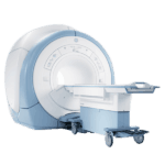
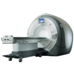
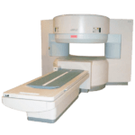
MRI scanning may actually be safe for patients with legacy pacemakers or implantable cardioverter-defibrillators
A new study was recently released that states MRI scanning may actually be safe for patients with legacy pacemakers or implantable cardioverter-defibrillators, as long as safety protocol is followed. Previously, these devices weren’t considered MRI-ready.
According to Saman Nazarian, MD, PhD, of University of Pennsylvania Perelman School of Medicine, in nine out of 2,103 scans (0.4%), eight of device resets to backup mode were transient. The device resets to a backup mode immediately after scanning and needs to be watched for, Nazarian said.
The reset couldn’t be fixed by device programming in one case, and this was only due to the fact that the patient’s legacy pacemaker had less than 1 month battery life left and the device needed to be replaced.
“If power-on reset occurs, the device reverts to an inhibited pacing mode. Therefore, in pacing-dependent patients, the device may transiently cease pacing because of electromagnetic interference, and electrocardiographic monitoring and pulse oximetry are warranted so that the scanning can be stopped if inhibition of pacing occurs,” the authors said. Their report was released online in the New England Journal of Medicine.
Safety protocol that was specified beforehand dictated that pacing be switched to asynchronous mode for pacing-dependent patients; pacing placed in demand mode for others; and tachyarrhythmia functions disabled.
Get Started
Request Pricing Today!
We’re here to help! Simply fill out the form to tell us a bit about your project. We’ll contact you to set up a conversation so we can discuss how we can best meet your needs. Thank you for considering us!
Great support & services
Save time and energy
Peace of mind
Risk reduction
In regards to the changes in device parameters, 1% of the patients had P-wave amplitude fall by at least 50% after having the MRI scan. There were no adverse reactions of the patients noted in the 63% that had a follow-up after 1 year. After that year, the most common changes described included decreases in P-wave amplitude (4%), increases in P-wave amplitude (4%), increases in atrial capture threshold (4%), increases in right ventricular capture threshold (4%), and increases in left ventricular capture threshold (3%).
The authors continued that “No change in device parameters that occurred either immediately after the MRI or at long-term follow-up in any patient was large enough to result in lead or system revision or device reprogramming.”
The complete study was made up of 1,509 patients. All completed 1.5T MRI scans that were deemed necessary by their physicians, despite all of them having a pacemaker (58%) or ICD (42%) that was not deemed MRI-ready by the FDA. The clinicians working on the study performed device interrogation at baseline and immediately following the MRI scan.
According to Nazarian’s group, “In our smaller study that was reported previously, we noted an association between thoracic imaging and changes in long-term right ventricular sensing and capture threshold. However, the current larger study, in which the follow-up period was longer, does not suggest any association between the region of imaging and detrimental changes in device parameters. The primary detrimental associations were a larger reduction in right atrial and right ventricular lead sensing immediately after the MRI with ICD systems than with pacemakers, as well as a larger reduction in long-term right ventricular lead sensing with longer lead length than with shorter lead length.”
The research and subsequent study was a single-center study, but the analysis included patients with a wide variety of cardiac devices. The number of each pacemaker or ICD was small, therefore a 20% rate of patients were lost to follow-up, which limited the studies full capabilities. However, the present findings still compliment those of the older, similar MagnaSafe Registry.


