CT Scanner Maintenance: Calibration

What are the advantages and the disadvantages of each modality and how they both can benefit any imaging department?
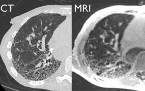
Both modalities have been around since the seventies, and they take axial, coronal, and sagittal planes of the human body. Then, 3D images are formed using computer algorithms to show images of bones, major organs and vessels of the heart and brain. But both modalities may not be suitable for all exams and patient population.
MRI machines come with different magnetic field strengths measured in Tesla, they can range from 1.5T all the way up to 7.0T on research machines. CT scanners come with different slice counts, from one single slice to 250+ slices. On MRI units the higher the magnetic field strength the more accurate and detailed the images are going to be. Likewise, the higher the slice count on the CT unit, the thinner the slices are, and the more detailed the final image is.
The big difference is how each modality acquires images during the exam. CT units use x-rays and MRI units use magnetic fields and radio frequencies to form 3D images.
How images are acquired on MRI Machines vs CT Scanners?
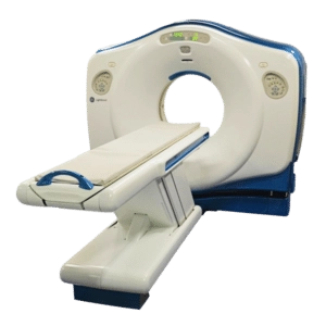
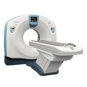
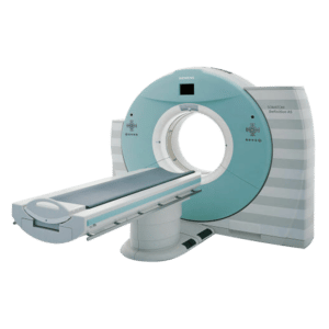
On a CT scan machine, a patient is placed on a table in a gantry for the exam. The X-ray tube rotates inside the gantry taking pictures of the body at different angles while the patient moves through the scanner. The images are formed by the different absorption rates of x-rays in human tissues. These differences are assigned a number that depict a certain level of darkness and lightness of the body part. For example, bone will be light on the image and soft tissues such as heart and lungs will be darker or a shade of gray.
MRI units use radio frequencies and strong magnetic fields to acquire the images. During an MRI scan, patients are placed inside the scanner with a coil placed over the body part being imaged. For example, there are different coils such as, head, extremity and neck coils. The coils enhance the signal of the MRI unit to produce a detailed image of the body part. The images are formed using the differences of water in the cells. For example, diseased or cancerous tissues contain more water in their cells than healthy tissues. MRI scanners use these differences called T1 and T2 weighted images, to produced detailed pictures of bones, ligaments and other soft tissue densities.
In both procedures, the images are reconstructed by a computer using mathematical algorithms to produce 3D images of bony and soft tissue anatomy. The advancement of both technologies has enabled the visualization of vascular structures in major organs such as the heart and brain during angiograms. Blood flow can be visualized during MR and CT angiography in real time. A fast diagnosis can be made of strokes and blood clots in emergent cases.
Get Started
Request Pricing Today!
We’re here to help! Simply fill out the form to tell us a bit about your project. We’ll contact you to set up a conversation so we can discuss how we can best meet your needs. Thank you for considering us!
Great support & services
Save time and energy
Peace of mind
Risk reduction
Advantages and disadvantages
MRI uses radio frequencies and magnetic fields to acquire images, which is the main advantage over CT exams, because it does not use radiation. However, patients with metallic devices such as pacemakers or prosthetic hips and others who suffer from claustrophobia are contraindicated for MRI exams. Ferromagnetic materials, such as IV pumps or O2 tanks pose a safety issue and cannot be in MRI scanning rooms.
CT uses radiation to acquire images. Metallic implants or the presence of ferromagnetic materials in the scan room are not issues for CT exams.
Which is the better? It depends on the patient population and the exams performed
CT machines use a rotating x-ray tube to acquire 3D images. CT exams are usually used to diagnose muscle or bone disorders, look for tumors, and diagnose lung and chest problems. Internal damage from accidents can be thoroughly examined by a CT Scanner as well, so they are frequently used in emergency rooms.
MRI uses radio frequencies and a magnetic field to do the same. MRI exams are better for soft tissue including tendon, ligament, and spinal columns, as well as diagnosis of brain tumors.
Each modality has something to offer which is why imaging departments should consider having both in their arsenal for different exams and patient populations. MRI’s price points depend on magnetic field strength or Tesla, CT’s price points are based on the slice count and generating power.
| MRI Machine | CT Scanner | |
| Imaging | Uses magnetic fields and radio frequency pulses to take images of internal organs. | Takes a series of X-ray “slices” of the body to produce a 3D image. |
| Length of Scan | From 10 minutes to 2 hours, depending on what looked for. | Usually within 5 minutes. |
| Radiation Risk | None. MRI machines do not pose a radiation risk because they do not use X-rays to produce their images. | About the same amount of radiation, one receives from background radiation in 3 to 5 years. |
| Patient Comfort/Risks | Painless and noninvasive. Some may be allergic to contrast dye. Claustrophobia risk and annoyance over long scan time. | Painless and noninvasive. Short scan time but radiation risk is present. |
| Limitation for Patients | Weight limit is usually 350 pounds. Patients with metal or cardiac implant are contradicted. | Weight limit is 450 pounds. Metals implants are okay for CT scans. |
| Average Cost | Starting from $150,000 and can got up to $1.2 million. | Starting from $65,000 on the lower slices, and up to $300,000 for the higher slices. |


