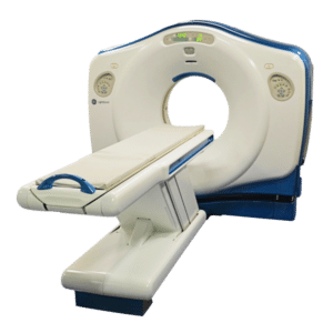
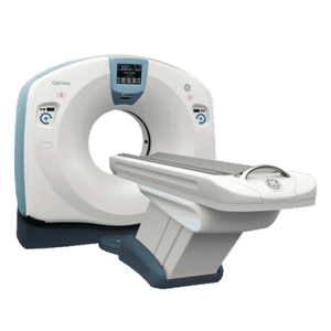
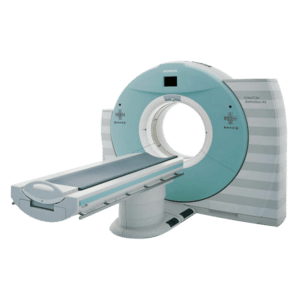
CT Scanner slices is determined by how many levels of anatomy are acquired in one rotation
CT scan machines come in single, dual source and multi-slice technology. CT Scanner slices is determined by how many levels of anatomy are acquired in one rotation. To be clear, 1st, 2nd and 3rd generation of CT scanners are single slice units, the later generations, fourth through seventh, are multi-slice units with slice counts going from 4 to 256
Single slice units have one x-ray tube and a single row of detectors. One slice is acquired in one rotation. By moving the patient through the scanner, the slices are acquired faster making volumetric scanning possible on a single slice unit.
Dual source CT units have two x-ray tubes and two sets of detector arrays. These units can simultaneously image a specific body part at two different angles. For example, DSCT unit can capture the phases of the cardiac cycle during systole and diastole in real time. This unit is ideal for Cardiac studies.
All multiple slice units are capable of doing, CT angiography, CT Fluro, etc. These scanners have multiple detector arrays and a single x-ray tube, they take multiple slices in one rotation. For example, in a 16 slice CT unit, 16 levels of anatomy are acquired in one rotation of x-ray tube and detector assembly.
The difference between these models is the speed at which the images are acquired. The more detectors the more slices acquired in one rotation. Multiple detector arrays increase slice counts in one rotation and reduce scan times.
Here is a little history on the development of the CT scanner slices
The first generation
Back in the 1970’s CT units used a single x-ray tube and detector.
The data acquisition was based on translate rotate principle. The single tube and detector rotated one degree at a time and took one picture. Next, the tube and detector moved another degree and took another image from a different direction and so on. This repeated for 180 degrees around the body part being imaged. The first-generation scanners took 4.5 to 5.5 minutes to image one slice.
This slice-by-slice scanning was time consuming which led to the idea of moving the patient through the CT unit while being scanned. The x-ray beam traces a path around the patient called beam geometry or helical CT. So instead of one slice being imaged a volume of tissue was acquired during the scan. These single slice scanners were called single-slice helical CT scanners or volume CT scanners.
The second generation
These single slice scanners employed the same principles as the first but with some structural differences such as 30 detectors and multi-pencil beam x-ray tubes. This fan-shaped beam translates across the patient to collect a set of transmission readings. After one translation, the tube and detector array rotate over a bigger area instead of one degree.
The third generation
These scanners uses the fan beam geometry but the array of detectors are arced 30 to 40 degrees to catch the attenuating x-ray beam. The path traced is circular vs. semi-circular which decreased the scan time as well.
Get Started
Request Pricing Today!
We’re here to help! Simply fill out the form to tell us a bit about your project. We’ll contact you to set up a conversation so we can discuss how we can best meet your needs. Thank you for considering us!
Great support & services
Save time and energy
Peace of mind
Risk reduction
The fourth generation
These machines employ a stationary ring of detectors. This single slice unit uses one x-ray tube that rotates inside a stationary ring of detectors inside the gantry.
In 1998, multi-slice CT units (MSCT) were developed that were based on multi-detector technology. In multi-slice units there is one x-ray tube and an array of detectors instead of a single row. A wider area could be covered in one rotation. For example, in a 256- slice unit there are 912 detectors on the transverse side x 256 detectors on the cranio-caudal side. The cone angle on the fan-shaped beam is about 120 degrees with a gantry rotation. This unit in one rotation will yield 256 slices compared with older models that acquired 64 slices or less with one rotation.
256 slice units are also ideal for cardiac studies. The whole heart can be imaged in one 360-degree rotation. Other multi-slice units are capable of doing cardiac studies but take longer to image the heart. The more slices the quicker the scan times and the better image quality.
To sum up, the current models of CT units take 3D images instead of one slice pictures per rotation. Whether it’s a single slice, 8 slice or all the way up to 256 slices units the patient moves through the gantry while being imaged. The increased slice counts and detector arrays in current CT units reduce scan times.
The differences between the single slice and multi-slice unit is the number of detectors inside the gantry. The detectors are arranged in an array in multi-slice units. Single slice units contain a single row of detectors inside the gantry. In multi-slice units the more detector arrays the more slices imaged in one rotation which means quicker scan times and better image quality.
Purchasing a multi-slice unit will depend on the studies being conducted, budget, patient population and space. Cardiac studies would require at least 32 slice counts to adequately image the heart valves and vessels at different phases of the cardiac cycle. All other studies such as Fluro CT, CT angiography , and CT Sim procedures can be done with lower slice counts.
As always, the price points are determined by slice count, bore size, generator power, and weight tolerances. Be sure to consult with your imaging team to determine which scanner will suit the needs of the department.
Sources:
Computed Tomography: Physical Principles, Clinical Applications, and Quality Control 4th Edition Euclid Seeram
https://en.wikipedia.org/wiki/Operation_of_computed_tomography


