What are the inner workings of a computerized tomography scanner (CT Scanner), sometimes called computerized axial tomography
In this article we will describe the inner workings of a computerized tomography scanner (CT Scanner), sometimes called computerized axial tomography or computer aided tomography (CAT scanner), and the types of exams that it can perform.
The origins of Computer Aided Tomography, who invented the first CT scanner?
The first CT scanner was invented by British engineer Godfrey Hounsfield of EMI laboratories. In 1971, Houndsfield along with Dr. Ambrose scanned a female patient that suffered from a brain tumor in Atkinson’s Morley Hospital. That first scan took days to image the tumor.
By 1973, the United States installed its first unit and by 1980, three million CT exams had been recorded.
How does a CT scanner work?
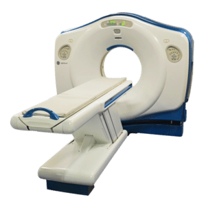
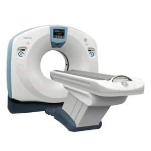
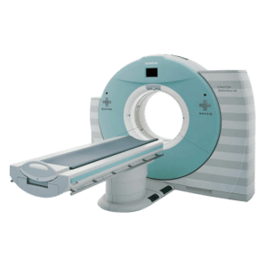
A basic CT machine consist of a gantry, a moveable table, and a control room. Inside the gantry contains the x-ray tube and an array of detectors. The 3-D rendered image is accomplished by rotating the x-ray tube in an ‘arc’ around the patient while their body moves through the gantry, which is the circular structure in the middle of the CT scanner.
The x-ray tube uses a fan shaped beam to take 2D images of the coronal, sagittal, and cross-section planes of the body. One revolution equals a slice and produces a 2D image of that slice or plane. Slices are collected in a computer and the scanning process continued until the desired number of slices are achieved.
Then, advance computer algorithms are used to reconstruct 3D images of those planes or slices. Slice thicknesses range from 1mm to 4 mm and are stacked to see a 3D image.
The fully rendered 3D images reveal detailed images of bones, soft tissues, and organs.
Here’s how CT units acquires images
Image reconstruction relies on different tissue densities that attenuate the x-ray beam at different energies. For example, x-rays will not penetrate bone or thicker body parts as easily as soft tissues such as muscles, ligaments, and cartilage.
Thicker body parts, such as bones will be lighter in value and softer tissues such as muscles, will be darker. Those value differences are assigned numeric units which are fed into a computer to reconstruct a 3D digitalized image of the body part. A contrast dye can sometimes be used to show certain structures more clearly.
CT images reveal details of bone, muscle, cartilage and other soft tissues and organs. The scan can determine the size and shape of the lesion or fracture deep inside the body. CT 3D images make it easier for physicians to find the location of the problem by allowing them to rotate the image in space or view slices in succession.
Get Started
Request Pricing Today!
We’re here to help! Simply fill out the form to tell us a bit about your project. We’ll contact you to set up a conversation so we can discuss how we can best meet your needs. Thank you for considering us!
Great support & services
Save time and energy
Peace of mind
Risk reduction
How are CT Scanners used?
CT exams are indispensable for all procedures including surgical, neurological, cardiac, and GI studies. A CT scan is ideal for trauma patients that need to remain immobile. The patient is positioned inside the scanner and planar images are taken from the side, front, and cross-sectional areas without moving the patient. Then the body part, such as the C-spine, will be seen in 3D.
Today’s CT scanners can take over 64 slices in one revolution and can render accurate 3D images in a matter of minutes. The thin slices reveal, tiny blood clots, small lesions, fractures and other diseases on soft tissues and organs.
CT scans are also used in surgical procedures. For instance, there are CT units set up in surgery for head trauma cases. The surgeon can operate on the patient in the O.R. while imaging the head during the surgery.
Biopsy studies, and radiation therapy planning are accomplished with CT simulator units as well. The details of the lesion are imaged with pinpoint accuracy and aids in biopsy needle placement and radiation therapy planning.
The state-of-the-art scanners can automatically position the patients inside the gantry which allows the technologist to perform other functions.
There are different types of CT scanners and exams
Some CT scanners are specifically designed for various procedures and patient types such as:
- Bariatric,
- CT simulation procedures,
- Cardiac,
- Spiral,
- and Perfusion exams.
Standard CT units can perform the above exams with some proficiency. However, if the institution specializes in certain procedures or serves specific patient populations it’s best to get a CT scanner that is designed for that procedure. For example, bariatric patients may not fit inside the gantry of a standard CT unit. Therefore, a bariatric scanner would be the better choice.
When it comes to purchasing a CT scan machine there are many brands and models to choose from. What affects the cost of most scanners, is size, speed and slice counts.
CT Scanners will continue to be used in the future.
CAT scans have come a long way from the 1970s from simple one slice units to multi slice units of today. They are used in hospitals throughout the world in a wide array of studies, procedures, and settings. The future of CT technology will continue to evolve and grow. And with the application of Artificial intelligence the role of CT technology will be a primary imaging tool of radiology.
If you would like to speak to one of Amber’s CT experts, contact us here today!
References:
https://www.fda.gov/radiation-emitting-products/medical-x-ray-imaging/what-computed-tomography
http://xrayphysics.com/ctsim.html
https://www.britannica.com/biography/Godfrey-Newbold-Hounsfield


