The Philips Gemini TF 16 Slice PET/CT Scanner is designed for detailed anatomical and functional imaging

Philips Gemini TF Overview
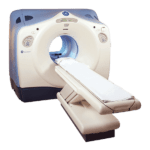
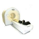
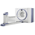
The Philips Gemini TF 16 Slice PET/CT Scanner is an advanced imaging system that combines positron emission tomography (PET) and computed tomography (CT) technologies. This hybrid system is designed for detailed anatomical and functional imaging, enhancing diagnostic accuracy for various medical conditions, particularly in oncology, cardiology, and neurology.
The Philips Gemini TF is a sophisticated imaging solution that combines the strengths of PET and CT technologies. It offers significant benefits, including enhanced diagnostic accuracy, improved patient care, and efficient workflow. With its advanced features like Time-of-Flight technology, high-resolution imaging, and user-friendly design, the Gemini TF 16 is a valuable tool for healthcare providers, contributing to better patient outcomes across various clinical applications.
Benefits:
Enhanced Diagnostic Accuracy: By integrating PET and CT, the Gemini TF 16 provides both metabolic and anatomical information, leading to more precise diagnoses.
Improved Patient Care: The high-resolution images allow for better disease detection and monitoring, facilitating timely and effective treatment planning.
Time Efficiency: The combined PET/CT scanning process reduces the need for multiple separate scans, saving time for both patients and healthcare providers.
Versatile Applications: The system is useful in a wide range of clinical applications, including oncology for tumor detection, cardiology for assessing heart function, and neurology for brain imaging.
Patient Comfort: The scanner is designed with patient comfort in mind, featuring a spacious gantry and fast scanning times.
Features:
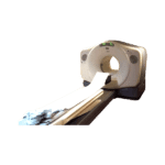
16-Slice CT Scanner: Provides high-resolution anatomical images and rapid acquisition times, enhancing diagnostic confidence.
Time-of-Flight (TOF) Technology: Improves image quality and lesion detectability by accurately measuring the time difference between photon detection events.
Advanced Reconstruction Algorithms: Enhance image clarity and reduce artifacts, leading to better diagnostic outcomes.
User-Friendly Interface: Simplifies operation and reduces the learning curve for technologists and radiologists.
Integrated Workstation: Allows for seamless data processing and image analysis, facilitating efficient workflow management.
Large Bore Design: Accommodates a wide range of patient sizes and improves patient comfort during the scanning process.